Back to All News

A complete toolkit for brain-tumor treatment

|
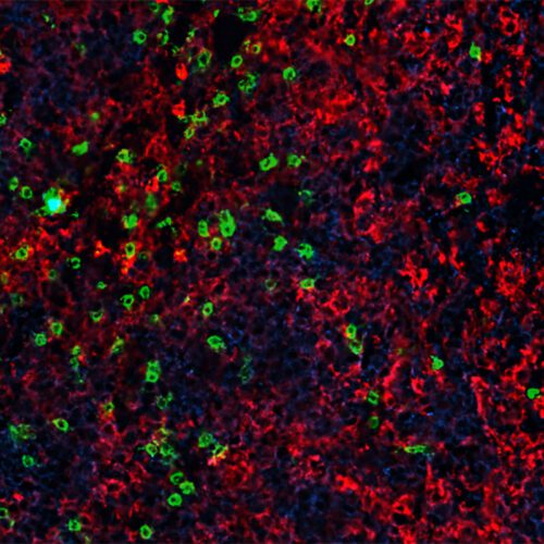
Human glioblastoma cells in culture: The markers for tumors, or nestins, are green; the nuclei are blue. Image courtesy of Albert... Human glioblastoma cells in culture: The markers for tumors, or nestins, are green; the nuclei are blue. Image courtesy of Albert Kim, MD, PhD

Every week, the brain tumor and neuro-oncology program at the Alvin J. Siteman Cancer Center at Barnes-Jewish Hospital and Washington University School of Medicine holds a tumor board meeting.

Attending are specialists in the field of brain-tumor diagnosis and treatment: neurosurgeons, neuro-oncologists, neuro-radiation oncologists, endocrinologists and neuro-intensivists, as well as representatives from psychology, neurology, genetics, hematology, pathology and nursing.

“The tumor board uses a collaborative approach to determine the best options for each patient with a brain tumor, whether benign, malignant or metastatic,” says Mary Spencer, executive director of neuroscience and orthopedic surgery. “Rather than relying on set protocols that may not prove effective, we tailor our patients’ care to their tumor characteristics.”

“Siteman’s program offers the most current surgical, medical and radiation therapies available, as well as opportunities to enroll in more than 35 clinical trials,” says Kaci Dannatt, oncology program manager. “More than 57 expert physicians, researchers and nurses specializing in treating brain tumors participate in the program.”

Targeting tumors, reducing risks

Washington University neurosurgeons at Barnes-Jewish Hospital have contributed to the success of the neuro-oncology program by using imaging technologies that improve the ability to see and remove tumors, while minimizing surgical risks to healthy brain tissue. Both functional magnetic resonance imaging (fMRI) and intraoperative MRI (iMRI) are part of this brain-mapping toolkit.

“Our neurosurgeons were among the first to use iMRI,” Spencer says. “Having worked on more than 1,600 cases, these specialists have gained considerable expertise in using iMRI for optimal resection of gliomas, pituitary skull-base tumors and spinal tumors, including metastases from lung and breast cancers.”

As a brain tumor is removed and cerebrospinal fluid drained, the initial fMRI becomes inaccurate for distinguishing tumor margins. Real-time iMRI gives neurosurgeons the updated information needed to complete the surgery. Siteman Cancer Center specialists also were among the first in the nation to use an MRI-guided, high-intensity laser probe to treat brain tumors that could not be removed with conventional surgery. In this procedure, heat from the laser kills cancer cells deep within the brain while minimizing damage to surrounding brain tissue, says neurosurgeon Eric Leuthardt, MD, who helped pioneer the minimally invasive laser procedure. The technique was first approved by the Food and Drug Administration (FDA) in 2009.

An unexpected result

Use of the laser probe recently led a team of neurosurgeons to a promising discovery: The technology can be used to penetrate the blood-brain barrier. This unexpected finding was made during a pilot study of 14 patients with glioblastomas who underwent minimally invasive laser surgery to treat the recurrence of their tumors.

“The laser killed tumor cells, which we anticipated,” says Leuthardt. “But, surprisingly, while reviewing MRI scans of our patients, we noticed changes near the former tumor site that looked consistent with the breakdown of the blood-brain barrier.”

In fact, Leuthardt and colleagues found that the laser treatment kept the blood-brain barrier open for approximately four to six weeks, providing a therapeutic window of opportunity to deliver chemotherapy drugs to the patients. Previous successful attempts by other groups to breach the barrier resulted in only modest benefits or were only successful for a short period of time—about 24 hours, not long enough for chemotherapy to be consistently delivered.

“Our finding is critical because most chemotherapy drugs can’t get past the brain’s protective barrier, which greatly limits treatment options for patients with brain tumors,” says Leuthardt, who also directs the Department of Neurosurgery’s Brain Laser Center and the Center for Innovation in Neuroscience and Technology.

“We are closely following patients in the trial. Early results indicate they are doing much better on average, in terms of survival and clinical outcomes, than what we would expect,” Leuthardt says. He notes the need for caution, however; additional patients need to be evaluated before conclusions can be drawn. Nevertheless, he adds, “we are hopeful this technology will open new avenues to treating these devastating brain tumors that cause great suffering for patients and their families.”

Expanding capabilities in radiation and oncology

The growing expertise of Washington University neurosurgeons in treating patients with brain tumors has been matched by expanding capabilities in neuro-radiation oncology and neuro-oncology. A string of firsts highlights the growth of these modalities at Siteman Cancer Center.

In 2014, Siteman Cancer Center introduced what remains the only proton-beam technology available in Missouri and one of the few units in the region. This technology allows radiation oncologists to control radiation beams by depth, shape and radiation dose, thus enabling treatment of solid tumors—near sensitive structures or tissues—which were once considered untreatable.

Another innovation, MRI-guided radiation therapy, was approved by the FDA in 2012. Siteman Cancer Center is the first center in the world to use this advanced cancer-treatment technology. The integrated system combines radiation treatments with a continuous-MRI system, allowing a radiation-oncology team to determine whether any subtle movements to the tumor or surrounding tissue alter the delivery of radiation. A patient’s treatment plan may be adjusted immediately if changes are noted.

Brain-tumor treatment has benefited from advances made in DNA sequencing, and the Elizabeth H. and James S. McDonnell III Genome Institute at Washington University is a world leader in the field. Currently, researchers are investigating the effectiveness of personalized vaccines for brain cancer.

“These are just a few examples of how research efforts have made significant contributions to the quality of care we can offer patients with brain tumors,” says Dannatt.