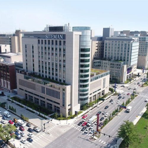
Siteman Flow Cytometry Core (SFC)

Overview

Siteman Cancer Center (SCC) Flow Cytometry (SFC) has expanded from nine fluorescent parameter analysis and sorting in 2001 to 27 color analysis and 22 color cell sorting in our current set of assets. We foresee experimental requirements expanding to beyond 40 colors with tools and techniques advancing at a fast pace. The general aims of SFC are to provide timely access to services, proficient flow cytometry expertise, and for staff to deliver the best possible operation and training. Given the varied academic origins of SFC customers, understanding and proficiency of flow cytometry are profoundly different from customer to customer. Depending on when a user learned flow within the span of the last 40 years, many changes of hardware and visualization standards have led to confusion and, worse, data interpretation inaccuracy. With this understanding, SFC aims have changed over the past several CCSG review periods to now include proper standardized instruction to users, which has already proven to increase the quality of data from basic research to clinical trial, and to mitigate expensive beginner failures.

Aim 1: Flow Cytometry analysis and cell sorting SFC provides reliable capacity for flow cytometry quantitative analysis in cancer research and exceptional cell sorting expertise with technical competency to cover all customer needs, including complex genetic sequencing purification and downstream recovery of highly multiplexed flow cytometry and mass cytometry developed target populations for cell sorting. SFC also provides access to machines for investigators who are proficiently trained to self-serve on a cell sorter so that they accommodate unpredictable patient schedules or other work outside of SFC’s normal staffing hours.

Aim 2: Flow Cytometry Instruction and experiment support SFC supports access to instrumentation with additional services that enhance the academic mission of the SCC. The cornerstone of SFC’s services involve instruction (from basic fundamentals to advanced expertise) of flow cytometry analysis and cell sorting. SFC supplies ongoing assistance with experiment design, data management, analysis, and data interpretation.

Aim 3: Developmental Interactivity Flow Cytometry is a very vendor driven science, with nomenclature and feature offerings typically driven by marketing exclusivity rather than the standardization that a science discipline craves. SFC management is involved in hardware, software and reagent development for several companies, benefitting SFC with reduced product pricing and ongoing development and support interaction. SFC is well informed with technology advances and can determine if new technology is appropriate for cancer center investigators well before launch.

SFC procures instrumentation from many companies with specialized strengths that comprise an extremely diverse list of services. SFC now has 10 self-service flow cytometry analyzers, including: seven three laser, 10 color FACScans and Gallios/Navios analyzers; two Attune NxT analyzers which are four laser, 14 color; and a Yeti five laser, 27 color analyzer. SFC offers staff operation on three cell sorters; a MoFlo four laser, 10 color sorter; and two Sony Synergy five laser, 22 color sorters. SFC also houses a self-service Aria SORP five laser, 18 color sorter. SFC intends to increase staff by one FTE in the next project period in anticipation of growing demand.

Leadership

Bill Eades

Self-service Flow Cytometry Analysis

The analysis section of the core offers self-service on four digital FACScans, updated to three lasers and 10 colors by Cytek Development, or two ThermoFisher Attune Nxt cytometers with four lasers and fourteen colors. For more information about optics and fluorophores, select the Self-Service Analysis page on the drop-down menu. 

Nine computer workstations are available in the core for post-acquisition data analysis at no charge. They are offered on a first-come, first-served basis and include FlowJo, Kaluza, Summit and WinList analysis suites. These applications are meant to be self-taught, but training is available by appointment.

Staff-operated Cell Sorting

Cell sorting is performed by a highly trained staff that is keenly aware of the physical science surrounding electrostatic sorting. Our staff members have over 50 years of collective experience from varied specialties,  including clinical process, sample preparation, and instrumentation design and support. All cell sorters are configured for flexibility. Given proper notice, the staff can set up for esoteric laser wavelengths and emission filter combinations, as well as up to four-way subset sorting and sorting into well plates. For more information about specific cell sorters, select the Cell Sorters page on the drop-down menu. 

As always, any pathogenic potential of samples must be disclosed, and an IBC protocol for your lab must be produced before any analysis acquisition or sorting will be scheduled.

Flow Cytometry Instruction

Instruction is required, regardless of previous experience, to operate any SFC instrumentation. A fundamentals module covers proper experiment design considerations as well as methods of analysis and interpretation. Subsequent modules are instrument operational software specific, and include acquisition of a mock sample and analysis instruction.

Instruction sessions are scheduled Tuesday, Wednesday and Thursday afternoons and start at 1PM. Capacity is limited and appointments are required. You may email [email protected] to schedule.

Location

Southwest Tower, Rooms 702, 702A, 703, 704

Pricing

Siteman member rates are for research and research associate members only. Inquiries regarding cancer center membership should be directed to the Siteman Cancer Center’s research administration office at 314-454-8110.

For a current fee schedule, see the drop-down menu at the top of the page.

Access

Call 314-362-9364 or e-mail [email protected]

Instrument scheduling calendars

NIH Public Access Policy

As of April 7, 2008, the NIH requires investigators with a publication using Siteman (or other NIH-funded) shared resources to submit (or have submitted for them) their final, peer reviewed manuscripts to PubMed Central(PMC) upon acceptance of publication, to be made publicly available within 12 months of publication. Many journals automatically submit these for authors, but Washington University also has assistance available through the Becker Medical Library. Please see https://becker.wustl.edu/services/author-analytics-and-support/compliance/#:~:text=The%20NIH%20Public%20Access%20Policy,upon%20acceptance%20of%20the%20publication for more information.

Publication Acknowledgment

If research supported by the Siteman Flow Cytometry results in publication, please acknowledge this support by including the following in your publication(s):

We thank the Alvin J. Siteman Cancer Center at Washington University School of Medicine and Barnes-Jewish Hospital in St. Louis, MO., for the use of the Siteman Flow Cytometry, which provided __________ service. The Siteman Cancer Center is supported in part by an NCI Cancer Center Support Grant #P30 CA091842.

Directions and Contact Information

Contact Information

Call 314-362-9364 or e-mail [email protected]

Directions to Siteman Flow Core Lab in the Southwest Tower of the Siteman Cancer Center

From the main Barnes Jewish Hospital entrance (and the elevated walkway from the underground Barnes parking garage, aka BJC South garage), follow the South Lobby main hallway past the South Cafeteria, turn left at the corridor to the Barnard elevators (or right if you are coming from the Wohl hospital entrance), and continue past the Barnard elevators to another foyer for the Southwest Tower elevators (signs should guide you to the elevators). (Occasionally the doors that separate the two foyers are closed and appear secure. You may open and walk through them at any time to get to the Southwest Tower.) Take the elevators up to the Seventh Floor and exit the elevator foyer towards the left hallway and glass double doors. These require a card swipe, so call 314.362.9364 with the adjacent phone, and we will come and get you. Office and Synergy/Attune instruments are in SWT702, the first door on the left, and digital FACScans, FACSAria, and MoFlo are in SWT703, the second door on the left.

We are not located in the CAM building.

NOTE: If coming from the Children’s Hospital parking garage, you can use the elevated walkway to go to the Wohl stairway, then take the first floor hallway to the Southwest Tower.

Current Fee Schedule

Cell Sorting or Analysis (core staff operated)
* Siteman CCSG member – $79.25/hour
Non-member – $110.50/hour

Flow Cytometric Analysis (self-service) low complexity (10 colors or less)
* Siteman CCSG member – $21.16/hour
Non-member – $31.68/hour
Charge for 15 minute increments

Flow Cytometric Analysis (self-service) high complexity (over 10 colors)
* Siteman CCSG member – $25.79/hour
Non-member – $36.31/hour
Charge for 15 minute increments

Self-Service Sorting
* Siteman CCSG member – $50.37/hour
Non-member – $53.93/hour

Instruction (data acquisition/analysis)
* Siteman CCSG member – $81.92/hour
Non-member – $127.99/hour

Live Cell Analysis
*Siteman CCSG member – $3.15/hour
Non-member – $3.36/hour

Cancellation Policy

The Siteman Flow Cytometry Core cancellation deadline is 24 hours before the scheduled start of any appointment.

Cancellations made after the 24 hour deadline are considered late and may incur full usage fees.  If someone else schedules and uses your cancelled time, you will not be charged.  If no one else schedules or uses the time slot, you will be charged full usage fees for your late cancellation.  No call, no shows also incur fees for the entire reservation.

For all sort appointments, users are charged from the beginning of their scheduled sort time.  Late arrivals will be charged for the time between the scheduled start time and actual arrival.


*Siteman member rates are for CCSG members only. Inquiries regarding cancer center membership should be directed to the Siteman Cancer Center research administration office at 314-454-8110.

Inquiries regarding flow cytometry services should be directed to 314-362-9364 or [email protected].

Self-Service Analysis

The analysis section of the core offers self-service on four digital FACScans, updated to three lasers and ten colors, or two Attune Nxt cytometers with four lasers and fourteen colors. Optics are fixed, and specifications/example fluorophores are as follows:

FACScans (10 color)

  • Blue laser (488nm excitation) FL1 (530nm emission centering wavelength/30nm bandwidth)- Alexa 488, FITC, GFP, YFP, CFSE
  • Blue FL2 (585/42)- PE, Propidium Iodide
  • Blue FL3 (620/20)- PE/Texas Red, ECD
  • Blue FL4 (695/40)- PerCP/Cy5.5, 7-AAD
  • Blue FL5 (740 LP)- PE/Cy7
  • Red laser (637nm) FL1 (660/20)- APC, Alexa 647
  • Red FL2 (725/20)- Alexa 700
  • Red FL3 (740 LP)- APC/Cy7, e780
  • Violet laser (407nm) FL1 (450/50)- Pacific Blue, Brilliant Violet 421
  • Violet FL2 (545/30)- Pacific Orange, Krome Orange, Live/Dead Yellow

Attune Nxt (14 color)

  • Blue laser (488nm excitation) BL1 (530nm emission centering wavelength/30nm bandwidth)- Alexa 488, FITC, GFP, YFP, CFSE, Live/Dead Green, DyeCycle Green, CellROX Green
  • BL2 (695/40)- PerCP/Cy5.5, PE/Cy5.5, PE/AF700, PerCP, 7-AAD
  • Red laser (637nm) FL1 (670/14)- APC, Alexa 647, SYTOX Red, FxCycle Far Red
  • RL2 (720/30)- Alexa 680, Alexa 700
  • RL3 (780/60)- APC/Cy7, e780, Live/Dead Near – IR, DyeCycle Ruby
  • Violet laser (405nm) VL1 (450/40)- Super Bright 436, Brilliant Violet 42, eFluor 450, Pacific Blue, BD V450, VioBlue
  • VL2 (525/50)- eFluor 506, Brilliant Violet 510, Pacific Green, BD HorizonV500, VioGreen
  • VL3 (610/20)- Super Bright 600, Brilliant Violet 605, Pacific Orange
  • VL4 (710/50)- Super Bright 645, Brilliant Violet 650
  • VL5 (660/20)- Super Bright 702, Brilliant Violet 711
  • VL6 (780/60)- Brilliant Violet 786
  • Yellow Laser (561nm) YL1 (585/16)- PE, Alexa 555, TdTomato, Propidium Iodide, SYTOX Orange, DyeCycle Orange
  • YL2 (620/15)- PE/Texas Red, PE/AF610, Live/Dead Red, mCherry, DsRed
  • YL3 (780/60)- PE/Cy7, PE/Vio770, DyeCycle Ruby

LINK:

Instrument scheduling calendars

Inquiries regarding flow cytometry services should be directed to 314-362-9364 or [email protected].

Cell Sorters

Beckman Coulter MoFlo (highly modified)

This cornerstone of the core allows for four detectors off a 200mw 488nm blue laser, up to three detectors off a 100mw 640nm red laser, and up to three detectors off a 100mw 405nm violet laser. A krypton-ion laser is also available for configurations involving 350nm (UV), 530nm (green), 568nm (yellow) and 752nm (NIR). Adequate notice is required for optics changes on the krypton laser as well as up to an hour allotted for the changeover time. A Propel Labs Co-Lase tower allows simultaneous use of four lasers and eleven emission detectors. The MoFlo is best known for the lowest cell sorting shear factor and small sample accuracy, as it has a manual sample station for minimal sample dead volume.

Sony iCyt Synergy BSC

The Sony SY3200 “Synergy” cell sorter is known for its very accurate high-speed sorting, pushing the Poisson limits for speed while maintaining excellent viability and purity results. In addition, unique reflective collection optics with over 375um laser intercept spacing and the newest “sputtered” hard coating emission filter technology have combined to attain new benchmarks for sensitivity and laser isolation. This instrument has two HAPS (highly automated parallel sorters), which can perform two different experiments simultaneously. Each HAPS module has 21 colors and five laser wavelengths: 355nm (UV), 405nm (violet), 488nm (blue), 561nm (yellow) and 640nm (red). The sorter modules are housed in a Class II biological safety cabinet, which has a unique passive aerosol evacuation feature that is as robust as the Baker hood itself. Also, each HAPS module is capable of four-way sorting and plate deposition.

BD Aria Ilu SORP

The Aria SORP has choices of 70um, 85um, 100um, and nozzles, and is equipped with an ACDU plate robot.  Laser wavelengths are 355nm, 405nm, 488nm, 561nm, and 640nm, and the sorter has 18 fluorescent detectors.


FAQs

Does the SFC prepare samples for sorting?

Our core can help plan and design experiments in regard to appropriate conjugate markers and proper process. We do not harvest, disassociate, or prepare flow cytometry samples for analysis or cell sorting. This is because of the sheer diversity of tissues and organisms dealt with, and we feel that there is mentorship available within your own specialty.

What cell types can I sort?

Non-adherent cells from culture, whole blood, PBMCs, bone marrow and cells from solid tissue are all acceptable samples for flow cytometry once disaggregated and red cells removed by lysis. Primary human samples or samples infected with BSL2 pathogens require special handling and notice must be given to SFC prior to sorting.

What cell concentration and sort tubes should my cells be in?

Single cells must be suspended at a density of 10-15 million cells/ml and passed through a 50um nylon mesh filter prior to sorting in order to reduce the chances of clogging the flow cytometer and its tubing. Cell concentration influences the rate of flow sorting, which can progress anywhere from 1,000–20,000 cells/second. Incorrect suspension density may lead to higher sort speeds and lower yield or recovery.

What size tubes do I need?

In general, cell suspensions for sorting should be brought in BD Falcon 5mL polypropylene round-bottom tubes (REF352063) with starting sample volumes no less than 300uL.

How many cells should I bring?

The number of cells to prepare for sorting varies greatly by experiment but is ultimately dependent on how many cells you need to recover, the frequency of the target population and % yield. To put expectations in real terms, you should prepare for the worst-case of 70% yield, which is apart from the idea of know aggregation gated by pulse width.  When you absolutely require a recovered number of cells for a downstream process, presort cell numbers should be 1.5 X target frequency.  Beginning with a surplus will assure that you will get what you need.

What nozzle size do I need?

When sorting, we try to maintain a ratio of 6:1, nozzle size to largest cell size in suspension. This will reduce shear factors and limit stresses put on the cells during the sorting process. While most non-adherent cell lines and cell suspensions will do fine with the 70um or 100um nozzle, other sensitive cells may require larger nozzles.  Please discuss with a member of the SFC if you are unsure what to request.

What sort of buffer should I use?

For a suspension buffer, we recommend Hank’s Balanced Salt Solution (HBSS) or Phosphate Buffer Saline (PBS) with no calcium or magnesium, and no phenol red. For cell nutrition, the use of Fetal Calf Serum (FCS) or Bovine Serum Albumen (BSA) is recommended depending on the cell line, and should be between one and two percent. The use of EDTA is optional based on the cell line and its aggregation characteristics, and normally 1mM is all that is necessary.

What type of receiving media can I use?

Receiving media varies by cell type and experiment but in general should contain twenty to forty percent FCS or BSA and antibiotics (pen/strep or gentamicin). After sorting, cells should be allowed to rest for at least thirty minutes before being spun down and re-suspended in their usual medium. We can also sort directly into lysis buffer, but please remember that sorted cells are contained within a droplet of sheath and not merely cells alone. Volume is added to the collection tubes at a rate of about 2.5mL per 1 million cells with our larger 100um tip and 1mL per million cells with the 70um tip.

Do I need controls?

For proper instrument set up, controls are needed every time an experiment is done. Unstained cells, along with positive and negative controls, are needed. Positive controls are used for compensation of fluorochrome spectral overlap. Remember that a fluorochrome’s emission is measured, not in a single detector, but in all the detectors being used in the experiment. Positive controls must have an adequate signal and percent positive to adjust settings. Negative controls should consist of unstained samples and fluorescence minus one (FMO) fluorochrome staining where applicable. FMO staining should include positive staining minus one of the fluorochromes. Compensation controls are critical to the determination of what we call positive or negative for a given marker in an experimen;, they are absolutely critical to the success of the experiment.

How many populations can I sort simultaneously?

All of the sorters are able to sort up to four populations at once. Four-way sorts must be collected into 5mL polypropylene round-bottom tubes or Eppendorf tubes, as there is a limit to the maximum distance we can separate the sort streams.

What do I need to do to schedule a sort?

For instrument scheduling, please email [email protected]. The following information is required at the time of your request:

  • Name, phone number and PI
  • Cell or tissue type
  • Approximate size of the largest cell in population and an estimate of the total number of presort cells
  • How the cells have been treated (transfected, transduced, stimulated)
  • Which fluorchromes or fluorescent proteins are being used
  • Downstream applications for the cells after they have been sorted
  • Number of populations to be recovered
  • Bulk or plate sorting required


Instrument Calendars

Please follow the links below to schedule time on our machines:

Analyzers

Sorters

Incucyte_S3